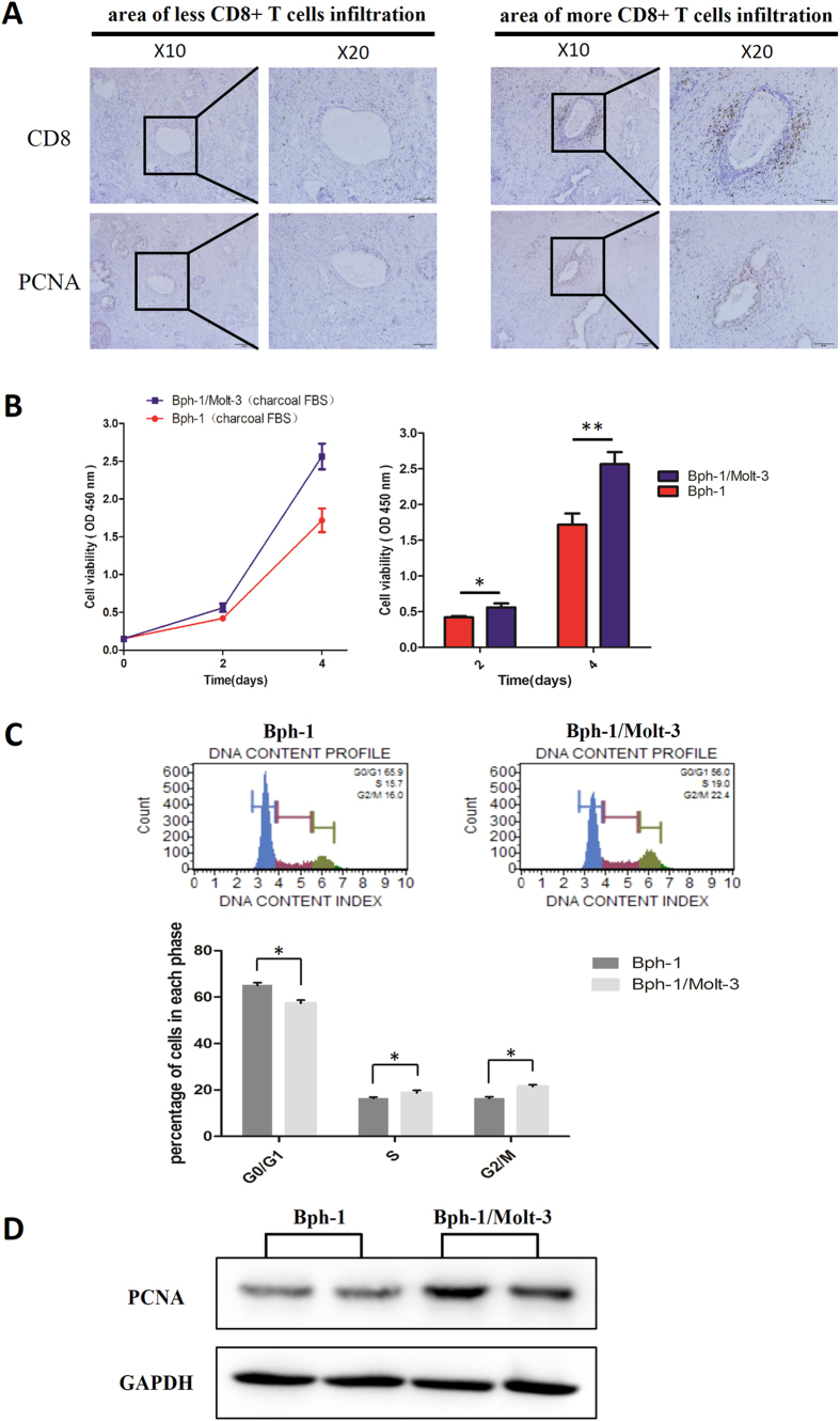
Figure 1. CD8+ T cells promoted the proliferation of BECs in the presence of low androgen.
(A) IHC staining in the serial paraffin sections for CD8 and PCNA from BPH patients who treated with Finasteride at least six months. The left panel is an area with less CD8+ T cells infiltration, and the right panel is an area with more CD8+ T cells infiltration; scale bar: 100 μm and 50 μm. (B–D): Bph-1 cells were co-cultured with/without Molt-3 cells in low androgen condition for 4 days. (B) Bph-1 cells were detected with CCK8 at days 2 and 4. Data are shown as the average OD value of Bph-1 cells and are mean ± SD. *P < 0.05, **P < 0.01. (C) Bph-1 cells were harvested for cell cycle assay at days 4. Data are shown as the percentage of cells in each phase and are mean ± SD. *P < 0.05. (D) The proteins of Bph-1 cells were harvested at day 4. Western blot assay was performed using an antibody for PCNA. GAPDH was used as a loading control (full-length blots were presented in Supplementary Figure 1).

