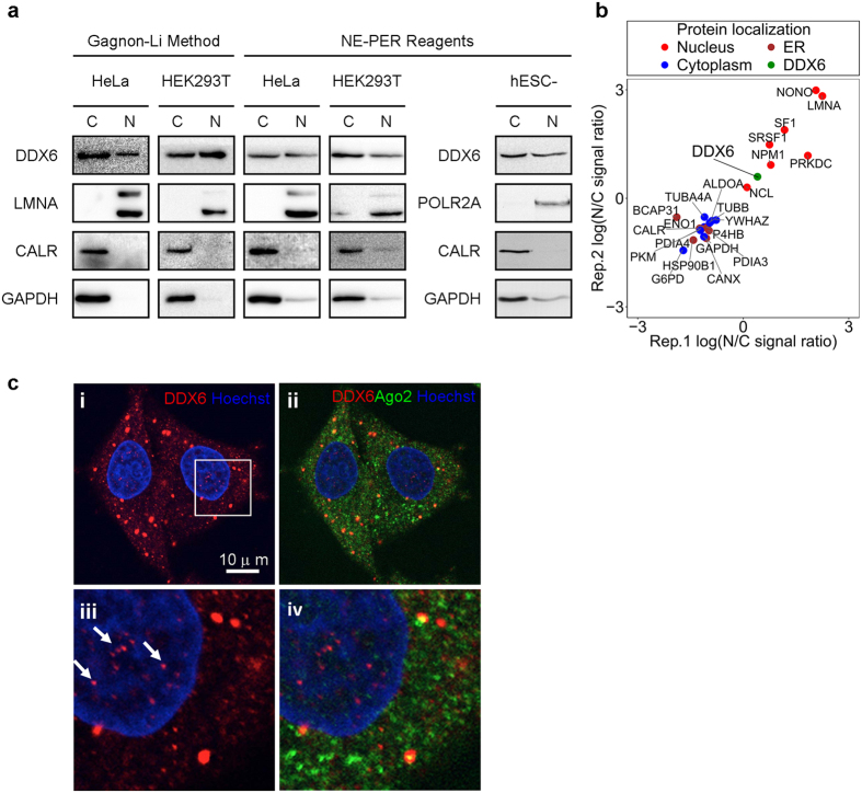
Figure 1. DDX6 is present in human cell nuclei.
(a) DDX6 is detectable in the nuclear extracts of human cell lines. Cytoplasmic and nuclear extracts from HeLa cells, HEK293T cells, and hESC-derived CM were prepared by NE-PER reagent or Gagnon-Li method. Equal amounts (proteins in μg) of nuclear and cytoplasmic extracts were separated by SDS-PAGE and analyzed by WB. LMNA and POLR2A p-Ser2: lamin A/C and phosphorylated RNA polymerase II subunit 1, nuclear markers; GAPDH: a cytosolic marker; CALR: an ER lumen marker. (b) Quantitative MS analysis confirms the nuclear presence of DDX6. Scatter plot showing the comparison of the protein nucleocytoplasmic signal ratios between the two replicates of the quantitative MS data. The original samples were fractionated by NE-PER to yield cytoplasmic and nuclear extracts as in (a). The N/C signal ratios were calculated using the summed-up extracted ion chromatogram (XIC) intensities, an estimate of the protein abundance. Several known cytosolic (blue), ER (brown), and nuclear (red) proteins were shown side-by-side with DDX6 (green). (c) IF detects endogenous DDX6 in the nuclei of human cells. HeLa cells were analyzed by immunofluorescence using antibodies against DDX6 (shown in red) and AGO2 (shown in green). Nuclei were stained with Hoechst 33342 (shown in blue) and images were digitally merged (c, i and ii). Bottom panels (c,iii and iv) display magnified views of the boxed areas in i. Arrows indicated endogenous DDX6 in the nuclei. Scale bar = 10 μm.

