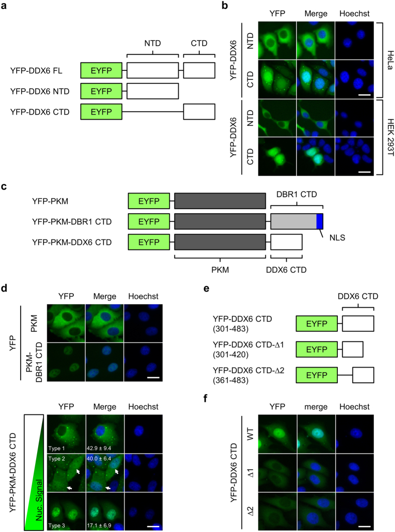
Figure 4. DDX6 CTD determines the nuclear localization of DDX6.
(a) Illustration depicts YFP-DDX6 FL, NTD, and CTD. NTD: the 1–300 AA; CTD: 301–483 AA. (b) YFP-DDX6 CTD localizes to the nuclei of human cells. HeLa and HEK293T cells expressing YFP-DDX6 NTD or YFP-DDX6 CTD were imaged with fluorescent microscope. (c) Illustration depicts the YFP-tagged chimeric proteins used for nuclear localization assay. PKM, a non-shuttling cytoplasmic enzyme, was used as the cargo for nuclear transport. ; DBR1 CTD, the CTD of a splicing factor DBR1, harbors a defined K/R-rich NLS, and was used as a positive control. DBR1 CTD and DDX6 CTD were fused to the C-terminus of YFP-PKM. (d) YFP-PKM-DDX6 CTD can localize to the nuclei. HeLa cells expressing YFP-PKM, YFP-PKM-DBR1-CTD, or YFP-PKM-DDX6 CTD were imaged 2 days after transfection. A phenotypic continuum of different nucleocytoplasmic distribution exists in the cell population expressing YFP-PKM-DDX6 CTD, and was roughly categorized into 3 types. Type 1: cytoplasm-dominant; type 2: equal cytoplasm/nucleus (indicated by white arrows); type 3: nucleus-dominant. Numbers represent means and standard deviations of the percentage of each type calculated from 3 replicates. (e) The diagram showing the truncation mutants of DDX6 CTD. DDX6 CTD was further truncated into CTD-Δ1 (301–420) and CTD-Δ2 (421–483) segments. White box represents DDX6 sequence. (f) Deletions in DDX6 CTD abolish the nuclear localization of DDX6. HeLa cells expressing YFP-DDX6 CTD, YFP-DDX6 CTD-Δ1 and YFP-DDX6 CTD-Δ2 were imaged with fluorescent microscope. (b,d and f) YFP signal is shown in green. Nuclei stained with Hoechst is shown in blue. Scale bar = 20 μm.

