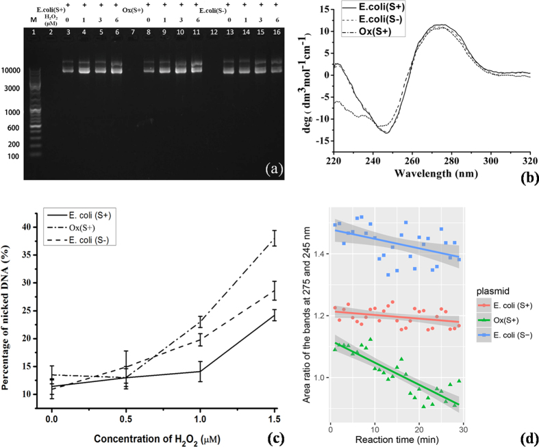
Figure 2. The in vitro antioxidant detections of the extracted plasmid DNA samples.
(a) The image of plasmid DNA gel electrophoresis, where the 63 μg/mL DNA samples treated with H2O2 up to 1.5 μM for 12 hours; (b) the circular dichroism spectra of the DNA samples; (c) the quantitated nicked DNA percentage in total amount of DNA, upon the oxidation; (d) the quantitated helicity upon the oxidative damage at 1.0 μM H2O2.

