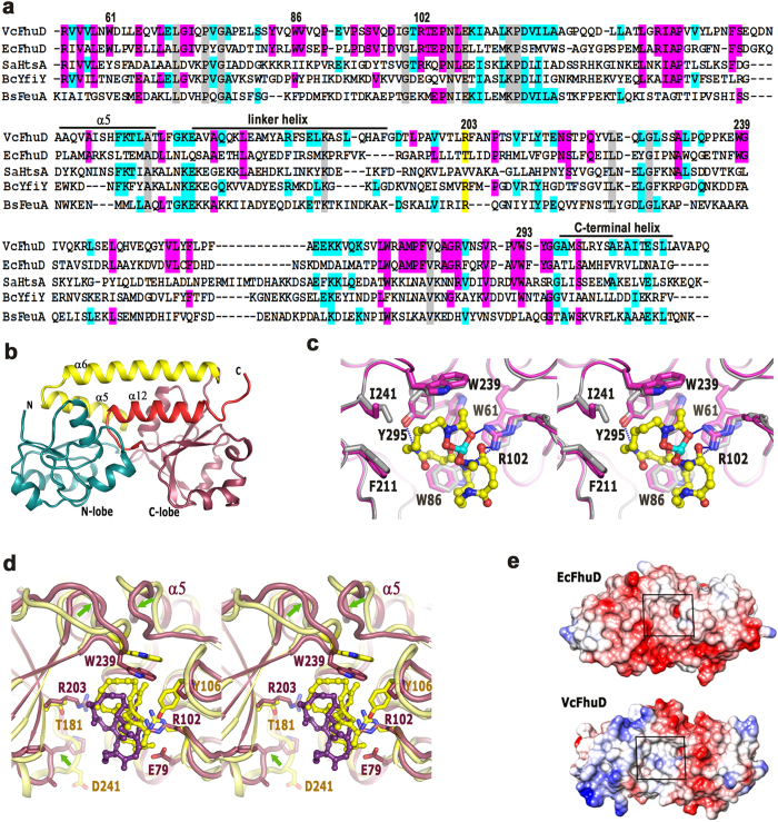
Figure 2.
(a) Sequence alignment of VcFhuD from V. cholerae, EcFhuD from E. coli, SaHtsa from S. aureus, BcYfiy from B. cereus and BsFeuA from B. subtilis. Numbering is based on VcFhuD sequence. Important motifs/residues are indicated by black bars and/or marked. Conserved residues are shown in gray. (b) Structure of apo-VcFhuD. (c) Stereo view of the superposition of holo and apo VcFhuD shown in magenta and white. The hydrophobic and polar interactions of VcFhuD with ferri-desferal are also shown here. (d) Stereo view of the comparison of ferri-desferal binding to VcFhuD (violet) and EcFhuD (yellow). Significant differences in loop conformation are shown by arrows. (e) Coulombic potential of EcFhuD and VcFhuD surfaces.

