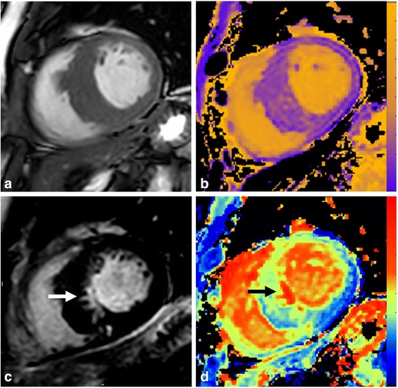
Fig. 1.

CMR images from a patient with asymmetric septal HCM. a SSFP imaging showing gross septal hypertrophy (>15 mm). b Native T1 map with colour scale ranging from 0 (purple) to 2000 ms (yellow). c Late gadolinium enhancement imaging showing a discrete area of replacement fibrosis in inferoseptum (white arrow). d ECV map ranging from 0 (blue) to 100% (red) confirming replacement fibrosis (black arrow)
