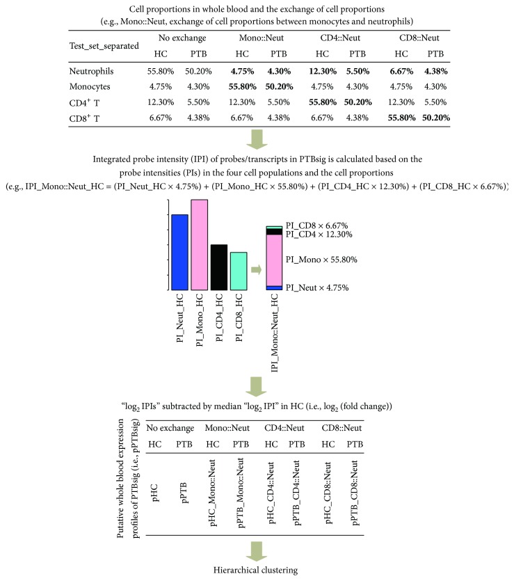
Figure 1.
Calculation of putative whole blood PTBsig (pPTBsig). Median cell proportions of the four separated cell populations in HC donors and PTB patients (i.e., neutrophils, monocytes, and CD4+ and CD8+ T cells) were obtained from Figure 3b of the published research [2]. The cell proportions were also artificially exchanged between neutrophils and the other three cell populations (e.g., Mono::Neut, exchange of cell proportions between monocytes and neutrophils). Then the integrated probe intensity (IPI) for each probe/transcript in PTBsig was calculated based on its probe intensity (PI) in the four cell populations and the (exchanged) cell proportions. Next, IPI was log2 transformed and subtracted from the median log2 IPI in HC donors. The output profiles demonstrated the relative expression levels (log2 transformed fold changes) of each probe/transcript in PTB patients compared to HC donors. pHC = pPTBsig based on the four cell populations from HC donors in Test_set_separated with no exchange of cell proportions. pHC_Mono::Neut = pPTBsig based on the four cell populations from HC donors in Test_set_separated with the exchange of cell proportions between neutrophils and monocytes. The other pPTBsig names were derived by similar abbreviation.

