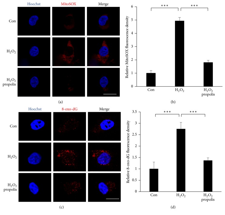
Figure 2.
Effects of methanol extracts of propolis on the H2O2-induced oxidative stress in SH-SY5Y cells. (a) Fluorescent images of MitoSOX Red signals in SH-SY5Y cells exposed to H2O2 for 1 h with or without propolis (10 μg/mL) for 2 h. Scale bar = 15 μm. (b) The quantitative analyses of MitoSOX Red signal intensity in (a). (c) Immunofluorescent CLMS images of 8-oxo-dG (red) with Hoechst-stained nuclei (blue) in SH-SY5Y cells exposed to 100 μM of H2O2 for 4 h with or without pretreatment with propolis (10 μg/mL) for 2 h. Scale bar = 10 μm. (d) The quantitative analyses of 8-oxo-dG immunofluorescence signal intensity in (c). The results are expressed as the mean ± SEM (n = 4 each), and the asterisks indicate a statistically significant difference from the indicated group value (∗∗∗p < 0.001).

