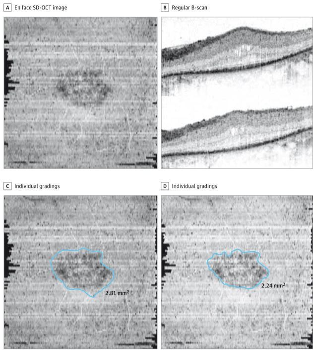
Figure 5. An Eye With a High Percentage Difference Between the Graders.
The difference between the graders was 23%. A and B, The precise boundaries of the preserved ellipsoid zone (EZ) band are difficult to discern on the en face spectral domain–optical coherence tomographic (SD-OCT) image and regular B-scans. C and D, The individual gradings are illustrated side by side. One of the graders who measured the preserved EZ area as 2.81 mm2 was not very precise in the grading; the other grader who measured the area as 2.24 mm2 was more meticulous.

