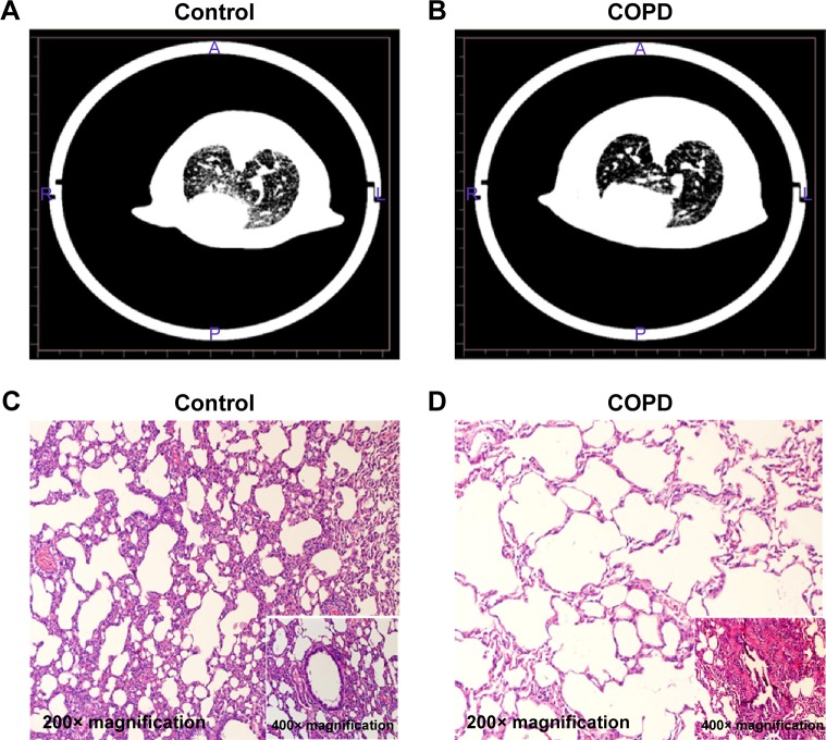
Figure 1.
CT analysis and histopathology examination of COPD rats.
Notes: The rats were treated with cigarette smoke and saline tracheal instillation for 90 days. Coronal CT images obtained in the control group (A) and in the COPD group (B). Histological examination of lungs in control-treated rats (C) and cigarette smoke-treated rats (D).

