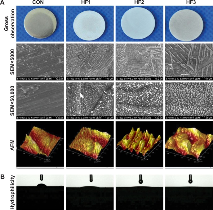
Figure 1.
Images of the titanium surface from gross observation, SEM, AFM and hydrophilcity.
Notes: (A) Micron grooves and corresponding nanoparticles on titanium surfaces were obtained under strict etching conditions and (B) hydrophilicity of the surfaces. Titanium surfaces were treated with 1% HF etched for 3 min, 0.5% HF etched for 12 min, and 1.5% HF etched for 12 min (respectively denoted as groups HF1, HF2, and HF3). Lower magnification of ×5,000, shows the overall microscale structures (upper images). Higher magnification of ×50,000 reveals nanoparticles (middle images). AFM of sample surfaces shows three-dimensional images and roughness of CON (control titanium surface) and treated groups (lower images).
Abbreviations: AFM, atomic force microscopy; HF, hydrofluoric acid; SEM, scanning electron microscopy.

