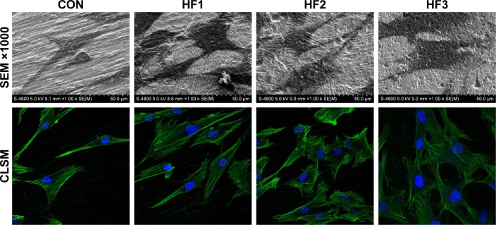
Figure 2.
hBMMSC adhesion and cytoskeleton were evaluated on the four surfaces after 3 days.
Notes: SEM images of hBMMSCs at magnifications of ×100 and ×1,000. Representative CLSM images of cells stained with DAPI to show the nuclei (blue) and FITC to show the actin filaments (green). CON represents control titanium surface; HF1, HF2, and HF3 represent cells, respectively, on the surfaces treated with 1% HF etched for 3 min, 0.5% HF etched for 12 min, and 1.5% HF etched for 12 min.
Abbreviations: hBMMSCs, human bone marrow-derived mesenchymal stem cells; SEM, scanning electron microscopy; CLSM, confocal laser scanning microscopy; DAPI, 4′,6′-diamidino-2-phenylindole; FITC, fluorescein isothiocyanate; HF, hydrofluoric acid.

