Figure 2.
Bactericidal activity of NDs.
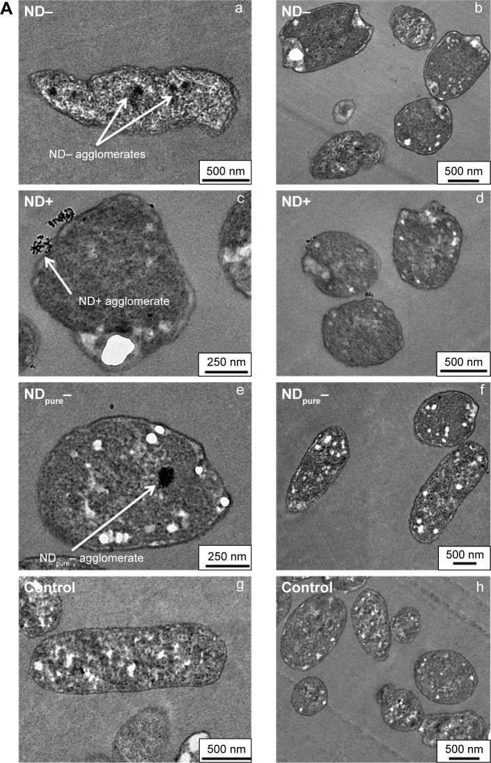
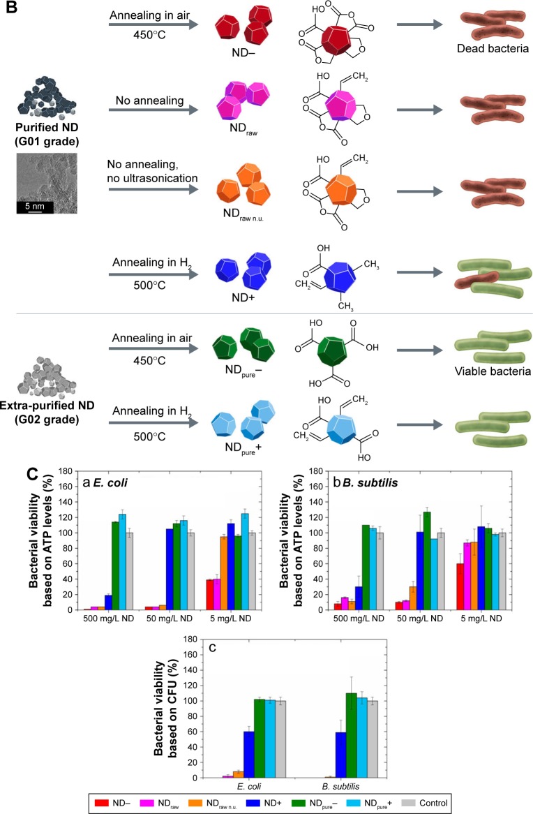
Notes: (A) TEM images indicate that, at sublethal ND concentrations of 0.5 mg/L, ND- is incorporated into E. coli cells and seems to deform the cellular shape (a, b); ND+ seems mainly to bind to cellular surface structures (c, d); Similar to ND-, agglomerates of negatively charged NDpure− are also found inside the cells, but they do not alter bacterial morphology (e, f); showing similar cell shapes to the ND-free control of E. coli (g, h). (B) Grades and pretreatments of NDs: a, negatively charged ND- and NDraw/NDraw n.u. were shown to exhibit strong antibacterial properties under aqueous conditions, while ND+ caused bacterial death only at high ND concentrations; b, NDpure, independent of their charge, did not show any bactericidal effects. (C) Antibacterial activity of NDs on E. coli and B. subtilis. (a,b) Negatively charged ND- and NDraw/NDraw n.u. strongly decreased bacterial viability measured by ATP levels in 15 min, while positively charged ND+ decrease ATP levels only at the highest ND concentrations for Gram-negative E. coli (a) and Gram-positive B. subtilis (b); (c) After incubation with 500 mg/L NDs, the determination of colony-forming units for E. coli and B. subtilis led to similar trends to the measurement of ATP, indicating that ND- and NDraw/NDraw n.u. are very effective at inhibiting bacterial growth, while positively charged ND+ are less bactericidal. Reprinted with permission from Wehling J, Dringen R, Zare R, Mass M, Rezwan K. Bactericidal activity of partially oxidized nanodiamonds. ACS Nano. 2014;8(6):6475–6483. Copyright 2014 American Chemical Society.111
Abbreviations: B. subtilis, Bacillus subtilis; CFU, colony-forming unit; E. coli, Escherichia coli; ND, nanodiamond; TEM, transmission electron microscopy; n.u, no ultrasonication.


