Figure 5.
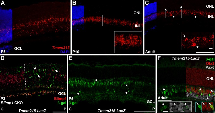
Tmem215-LacZ mimics Tmem215 expression in the retina. (A–C) Tmem215 in situ hybridization (red) in wild-type retinas. Nuclear counterstaining with DAPI (blue) is cropped for clarity. (A) Tmem215 signal in the central P5 retina is localized to the area of the retina containing bipolar cells and amacrines. (B) By P10, Tmem215 signal is strongly localized to the INL (inset). (C) In the adult retina, Tmem215 clusters signal in the outer INL (arrows, inset) suggest bipolar cell staining while less frequent clusters in the inner INL indicate amacrine identity (arrowheads, inset). (D–F) Tmem215-LacZ knock-in mice stained for β-galactosidase (green). (D) Postnatal day 2 Blimp1 CKO::Tmem215-LacZ transgenic retina. Central (C) is to the left while peripheral (P) is right. Blimp1 immunostaining (red) shows substantial deletion right of the dotted line. There are more β-gal+ cells (arrows) in the Blimp1 deleted peripheral region, mimicking the Tmem215 in situ data (Fig. 2). None of the β-gal+ cells in any region coexpresses Blimp1. (E) In P5 Tmem215-LacZ heterozygous mice, β-gal+ cells are localized in the future INL and primarily show bipolar cell morphology (arrows). A few cells with amacrine morphology are seen (arrowheads) and there is a central-to-peripheral gradient of β-gal expression, mimicking the normal developmental progression of bipolar cell genesis. (F) In adults, β-gal+ cells are localized to the INL. Most of the β-gal+ cells coexpress Otx2 (red) (arrows, blue insets) while a smaller population coexpresses Pax6 (gray, arrowheads, white insets). The Tmem215-LacZ transgene closely matches the Tmem215 pattern. Scale bars: (A–E) 100 μm for panels and 10 μm for insets; (F) 50 μm for the panel and 10 μm for insets.

