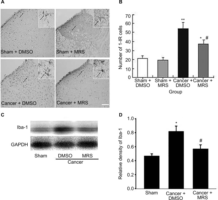
Figure 3.
Activation of microglia in the spinal dorsal horn induced by bone cancer and the inhibitory action of MRS2395.
Notes: (A) The staining of Iba-1-IR cells has been carried out for the sham + DMSO, sham + MRS2395, cancer + DMSO, and cancer + MRS2395 groups. The sham groups showed the morphology of lighter staining and more ramifications, suggesting inactivated microglia. The cancer + DMSO group showed deeper staining of Iba-1 and less ramification, suggesting activated microglia, and this activation was inhibited by MRS2395. (B) Comparison of average number of activated microglia in all groups. (C) Gel panels show products from the L4–L6 spinal cord taken from sham rats and bone cancer rats 9 days after administering with DMSO, MRS2395 using the Western blot. GAPDH was used as loading control. (D) Averaged data of immune blot densitometry showed that the relative level of Iba-1 protein, which was normalized to GAPDH, increased in bone cancer rats compared to sham + DMSO and sham + MRS2395 rats and partially suppressed by MRS2395. *P<0.05, **P<0.01 vs sham group and #P<0.05.
Abbreviations: Iba-1, ionized calcium-binding adapter molecule 1; Iba-1-IR, Iba-1-immunoreactive; GAPDH, glyceraldehyde-3-phosphate dehydrogenase; DMSO, dimethylsulfoxide; MRS, 2,2-Dimethyl-propionic acid 3-(2-chloro-6-methylaminopurin-9-yl)-2-(2,2-dimethyl-propionyloxymethyl)-propyl ester.

