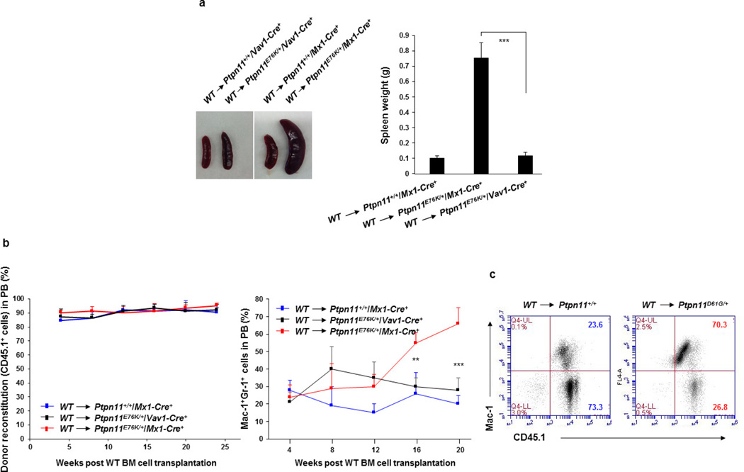
Extended Data Figure 3. Donor-cell-derived MPN is developed in Ptpn11E76K/+Mx1-Cre+ broad knock-in mice and Ptpn11D61G/+ global knock-in mice, but not Ptpn11E76K/+Vav1-Cre+ haematopoietic cell-specific knock-in mice transplanted with wild-type BM cells.
BM cells (2 × 10⁶) freshly collected from wild-type BoyJ mice (CD45.1+) were transplanted into lethally irradiated (1,100 cGy) Ptpn11E76K/+Mx1-Cre+, Ptpn11+/+Mx1-Cre+ (8 weeks after pI–pC administration), and Ptpn11E76K/+Vav1-Cre+ mice at 16-week old (CD45.2+). Spleen weights (n = 5 mice per group) (a), percentages of donor cell (CD45.1+) reconstitution (n = 8 mice per group) and percentages of donor cell-derived myeloid (Mac-1+Gr-1+) cells (n = 8 mice per group) in the peripheral blood of the recipients (b) were determined at the indicated time points following the transplantation. c, BM cells (1 × 10⁶) freshly collected from wild-type BoyJ mice (CD45.1+) were transplanted into lethally irradiated 3–4-month old Ptpn11D61G/+ and Ptpn11+/+ (CD45.2+) mice (n = 14 and 17 mice, respectively). Recipients were monitored for MPN development for 8 months. Percentages of donor cell (CD45.1+)-derived Mac-1+ myeloid cells in the peripheral blood of recipients were determined. Representative results are shown. Data shown in a, b are mean ± s.d. of all mice examined; **P < 0.01; ***P < 0.001. Source Data for this figure are available online.

