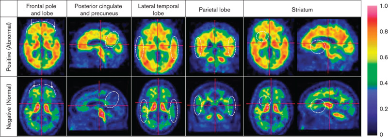
Fig. 1.

The electronic training programme (ETP) teaches the persons being trained to make a positive scan classification upon identification of several features in the following regions. Frontal pole and lobe: the lack of a marked sulcal pattern (dotted lines) and/or sharp intensity gradient from grey matter to cerebrospinal fluid. Posterior cingulate and precuneus: presence of cortical uptake in the circled region. Lateral temporal lobe: heightened uptake throughout and loss of the gyral/sulcal pattern (dotted circles). Parietal lobe: high uptake and decreased sulcal pattern within the dotted circles. Striatum: >50% uptake in the dotted region between the thalamus and the frontal lobe (axial or sagittal view).
