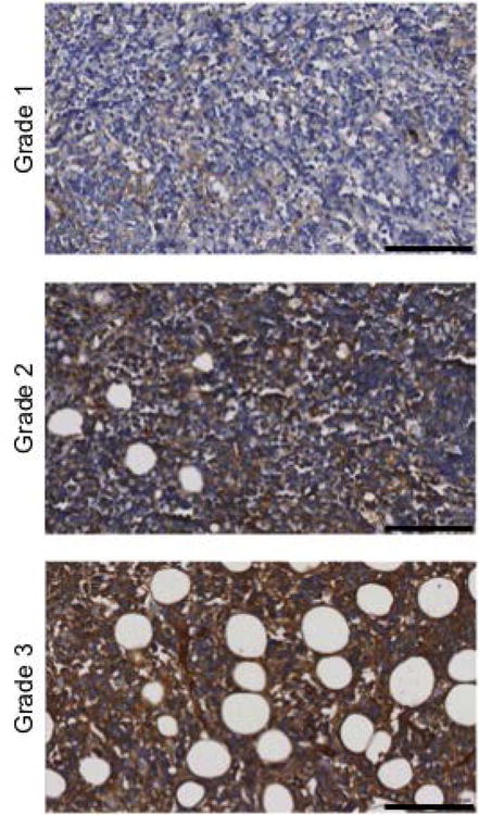
Figure 1. rVAR2 staining of Burkitt lymphoma tissue.

Presence of ofCS in BL tissues was analyzed by rVAR2 staining using the Ventana Discovery Platform. rVAR2 staining intensity was scored as negative (grade 0), weak (grade 1), moderate (grade 2), or strong (grade 3). Pictures are representatives of differentially scored tissues of grade 1-3. Staining intensities of ≥2 were considerate positive. The scale bars represent 100μm.
