
Figure 4. VAR2CSA binds to ofCS on Burkitt lymphoma cell lines.
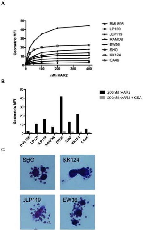
(A) rVAR2 binding to eight different BL cell lines. Signal-to-noise ration of the geometric mean fluorescence intensity (MFI) was tested using an anti-penta his alexa 488 antibody and flow cytometry. The graph is a representative of three independent experiments. (B) rVAR2 binding to BL cell lines with or without the co-incubation of 400 μg/ml soluble CSA in flow cytometry. (C) Binding of VAR2CSA-expressing P. falciparum infected erythrocytes to the BL cell lines as shown by microscopy after Giemsa staining.