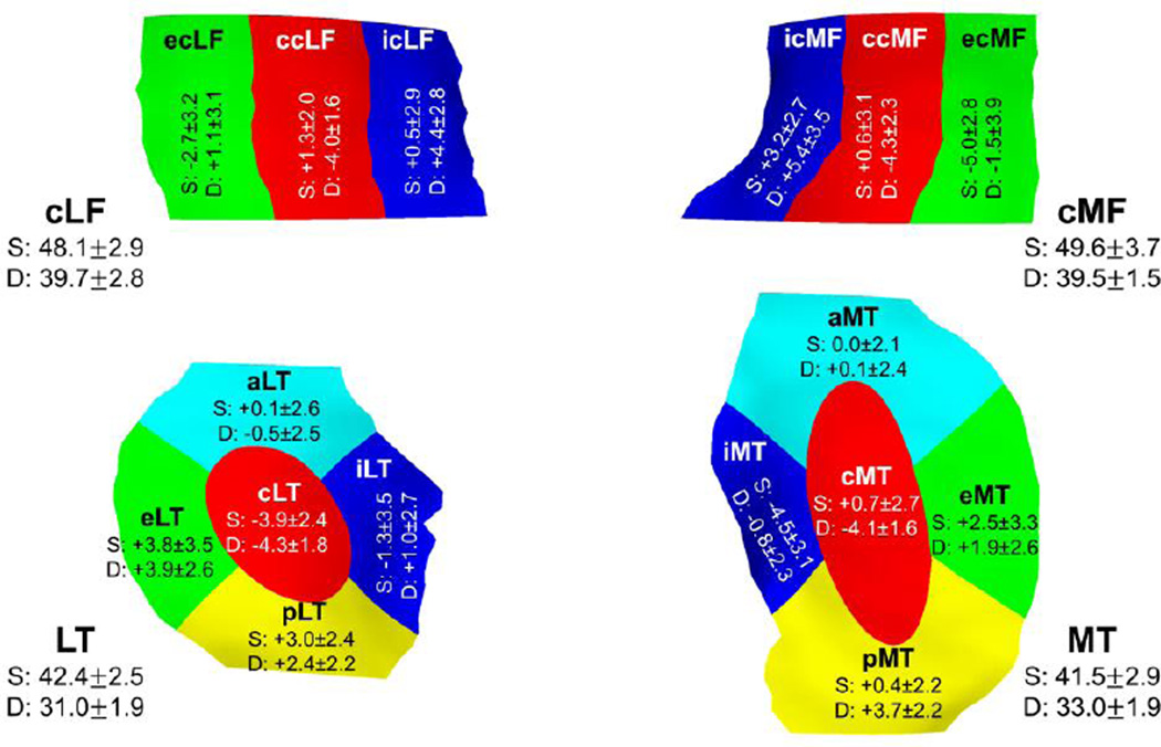
Fig. 2.
Difference between laminar subregional cartilage T2 times (mean ± SD difference in ms) and the cartilage T2 times in the superficial (S) and deep (D) layers of the respective total cartilages (mean ± SD in ms).The 3D illustration also shows the central (c), external (e), and internal (i) subregions of the central part of the medial (cMF) and lateral (cLF) femur and the central (c), external (e), internal (i), anterior (a), and posterior (p) subregions of the medial (MT) and lateral (LT) tibia.

