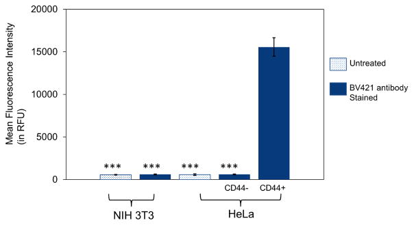
Figure 2.
Flow cytometry analysis using anti-CD44-BV421 antibody labeling to determine the CD44 receptor expression levels in CD44+ HeLa cells, CD44− HeLa cells, and CD44− NIH 3T3 fibroblasts. The CD44 receptor expression levels were determined by plating 75,000 cells per well in 24-well plates and incubating for 24 h before the experiment. Cells were then stained with CD44 antibody (mouse anti-human-CD44 antibody labeled with BV421) at a concentration of 1μg/mL in DMEM media for 30 min. After incubation, the spent media was removed and the cells were washed 3 times with PBS before trypsinization. The cells were then collected and analyzed using a BD FACS Aria III flow cytometer. Data points represent group mean ± SD (n = 6; ***P < 0.005; ANOVA).

