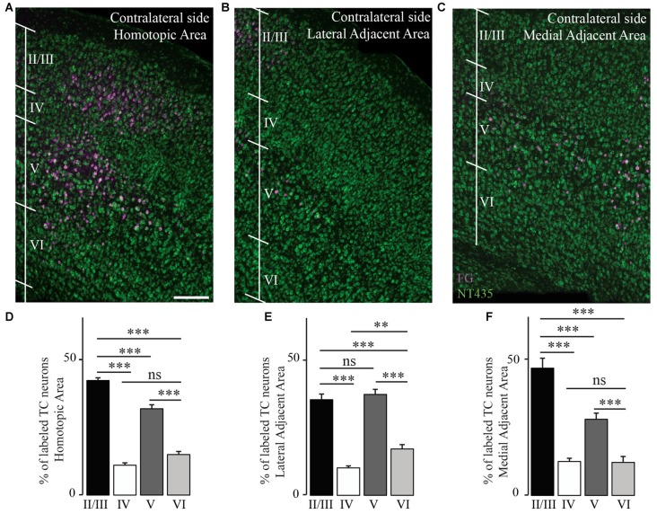
Figure 5.
Transcallosal projection neurons are primarily located in layer II/III and layer V of the contralateral cortex. (A) Confocal image of the homotopic cortical area (HA) showing the presence of labeled transcallosal projection neurons in layer II/III and layer V. (B) Confocal image of the cortex focusing on the lateral adjacent projection area (LAA) showing similar numbers of transcallosal projection neurons in layer II/III and layer V. (C) Confocal image of the cortex focussing on the medial lateral adjacent projection area (MAA) showing the location of transcallosal projection neurons in layer II/III and layer V. (D) Quantification of the repartition of transcallosal projection neurons across different cortical layers in the HA. (E) Quantification of the repartition of transcallosal projection neurons across different cortical layers in LAA. (F) Quantification of the repartition of transcallosal projection neurons across different cortical layers in MAA. Scale bars equal 200 μm in (A–C). One-way-Anova followed by Tukey post hoc test: ***p < 0.001; **p < 0.01.

