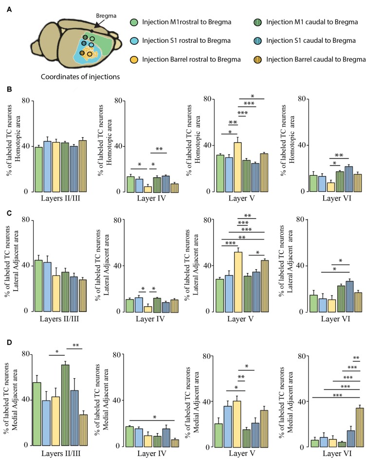
Figure 6.
Comparison of layer-specific organization of transcallosal projection neurons across the mouse cortex. (A) Scheme of the stereotactic coordinates used for retrograde tracing in the study. (B) Quantification of the location of transcallosal neurons in different cortical layers of the HA following injections in the cortex rostral to Bregma (full bars) and caudal to Bregma (dashed bars). (C) Quantification of the location of transcallosal neurons in different cortical layers in the LAA following injections in the cortex rostral to Bregma (full bars) and caudal to Bregma (dashed bars). (D) Quantification of location of transcallosal neurons in different cortical layers in the MAA following injections in the cortex rostral to Bregma (full bars) and caudal to Bregma (dashed bars). Green: injection in the motor cortex, blue: injection in the somatosensory cortex and yellow: injection in the barrel cortex. Two way- Anova (variables: (i) Areas of labeling and (ii) Injection sites) followed by Bonferroni post hoc test performed for each independent dataset. ***p < 0.001; **p < 0.01; *p < 0.05.

