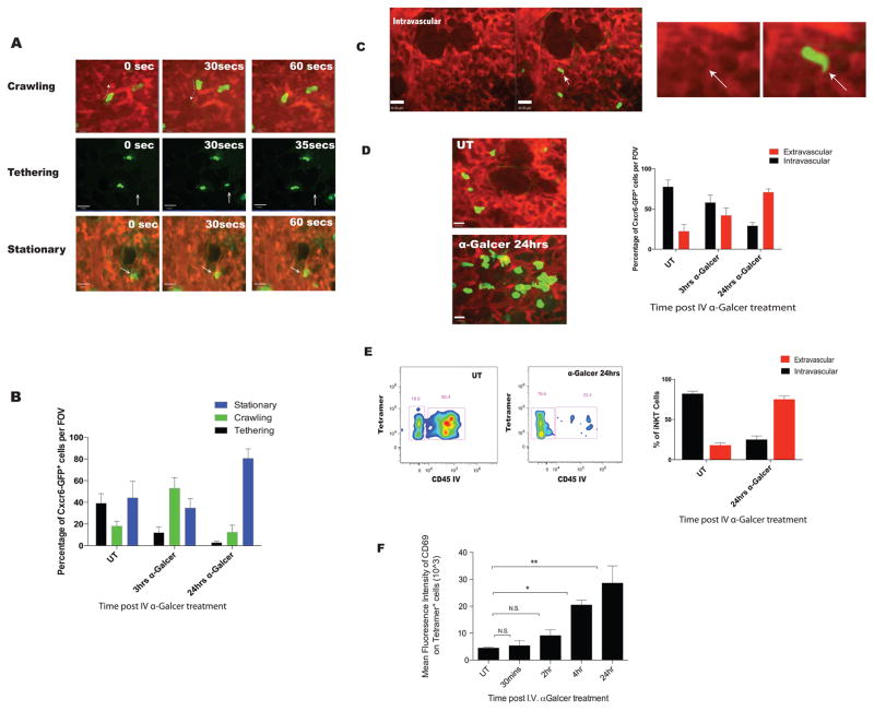
Figure 1. iNKT cells reside predominately in the lung vasculature migrating out upon stimulation with intravenous α-Galcer.
A. Three behaviours were observed for CXCR6GFP+ cells, identified by IVM in the lungs of untreated mice: (1) Crawling- movement of cells back and forth, (2) tethering- rapid movement of cells with blood flow with short 10–20 second stops and re-entering into blood flow and (3) stationary-cells shows protrusion, slight movement but the cell stays in the general area for 1 mins or longer. Green: CXCR6GFP+; Red: TRITC-dextran. B. Quantification of cell behaviors in vehicle treated (UT) or mice treated with 2μg of α-Galcer intravenously. C. Localization of cells was assessed using TRITC-dextran shadowing. Arrow indicates shadow formed by CXCR6GFP+ cells. D. Localization of CXCR6GFP+ cells was assessed in naïve mice (UT) or mice treated with treated with 2ug of α-Galcer intravenously. Extravascular CXCR6GFP+ take on a larger rounded phenotype and do not display shadows. E. Flow cytometry was used to assess cell localization of CD1d tetramer positive cells. “CD45 IV” are indicative of cells that are intravascular. Low to negative CD45 IV is indicative of extravascular localization. F. Dynamics of pulmonary iNKT cell activation based on mean fluorescence intensity of CD69 staining on tetramer positive cells after intravenous α-Galcer treatment. Error bars represent standard error of mean. ‘*’ and ‘**’ represent P < 0.05 and P<0.01, respectively. N = 3–5 animals per group.

