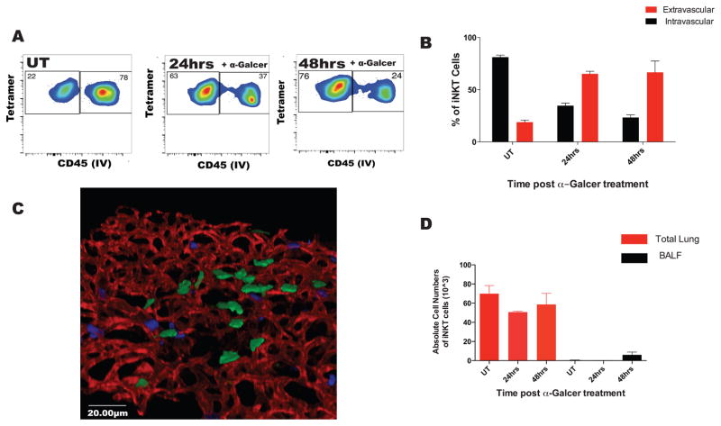
Figure 2. iNKT cells predominately migrate to the interstitial space after intratracheal glycolipid stimulation.
A. Mice were treated with aerosolized (400ng) α-Galcer. iNKT cell localization was analyzed by flow cytometry using anti-CD45 i.v injection. B. Localization of iNKT was quantified as in (1E). C. Lungs of mice treated for 48 hours with aerosolized α-Galcer were removed and whole mount staining was performed. 3D reconstruction was performed to assess spatial localization of CCXR6GFP+ cells after treatment. Green: CXCR6GFP+ cells; Red: CD31 stained endothelial cell; Blue: Ly6G stained neutrophils. D. The absolute cell counts for iNKT cells in the total lung homogenate and the bronchial-alveolar lavage fluid, BALF. Error bars represent standard error of mean. N = 3–5 animals per group.

