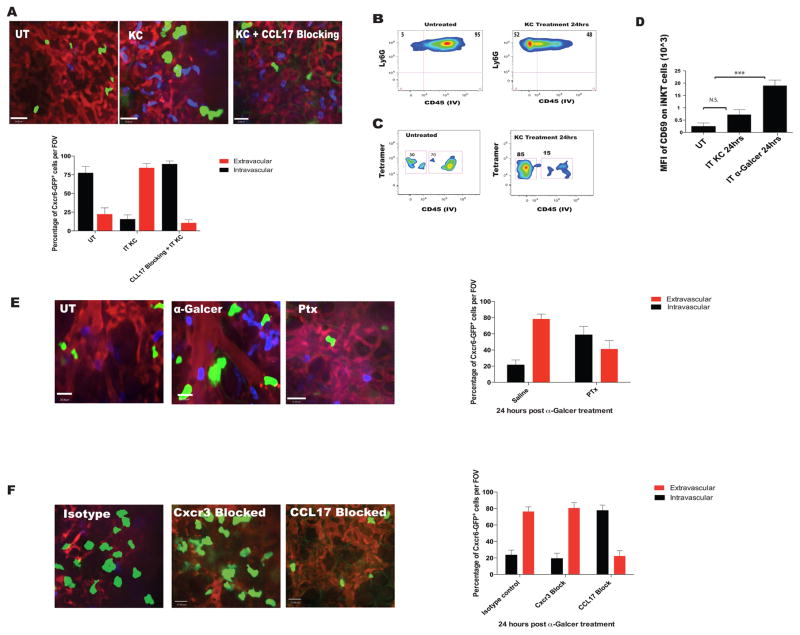
Figure 4. Pulmonary iNKT cell extravasate to the lung interstium in a neutrophil and CCL17 chemokine dependent manner.
A. IVM images and quantification of CXC6GFP+ cell localization based shadowing technique. Mice were treated with aerosolized KC (200ng) only or intraperitoneal CCL17 neutralizing antibody 30 minutes prior to KC. B and C. Neutrophil and iNKT cell localization based on CD45 IV staining after KC administration as in (1E). D. Mean fluorescence intensity of CD69 on tetramer+ iNKT cells after treatment with aerosolized KC; α-Galcer treatment (aerosolized) served as positive control. E. IVM was used to assess and quantify pulmonary CXCR6GFP+ cell localization in mice pretreated with pertussis toxin or saline control followed by i.v. α-Galcer for 15–24 hours. F. IVM was used to assess and quantify pulmonary CXCR6GFP+ cell localization in mice pretreated with CCL17 neutralizing antibody, or anti-CXCR3 antibodies, or isotype control followed by i.v. α-Galcer for 15–24 hours. Error bars represent standard error of mean. ‘***’ represent P<0.005. N = 3–5 animals per group.

