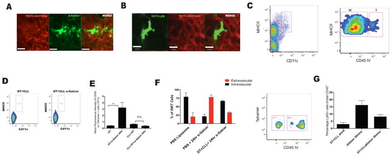
Figure 5. Dendritic cells residing in an extravascular position are the major cell type to take up α-Galcer and are crucial to iNKT cell activation.
A. Mice were injected i.v with the probe β-Galcer for 12–18 hours prior to imaging. IVM image of a β-Galcer+ cell. B. IVM image of a CD11c+ DC in the lung. C. Flow cytometry and anti-CD45 infusion was used to determine localization of CD11c+ MHCIIhi cells. D. CD11c-DTR mice were depleted of monocytes and CD11c+ DCs using i.v administration of CLL (200μL) and intraperitoneal administration of DT 24 hours prior to α-Galcer treatment. E. Activation level of iNKT cells was assessed based on mean fluorescence intensity of CD69 on tetramer positive cells. F. Localization of iNKT cells based on CD45 IV staining as in (1E). G. Neutrophil influx in the lung 30 minutes after α-Galcer treatment in mice depleted of monocytes and dendritic cells. Error bars represent standard error of mean. ‘**’ represents P<0.01. N = 3–5 animals per group.

