
Fig. 4.
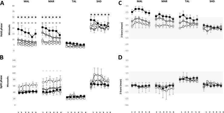
Mean ± standard error of mean of electromyographic activity (a, b) for right and left masseter (MAL and MAR); anterior temporalis (TAL) and suprahyoid (SHD) muscle in the mucosal anesthesia, incisal anesthesia, block anesthesia and the reference group during the hold phase (a) and split phase (b). The asterisk denote significant difference in the incisal anesthesia (white) and block anesthesia (black) compared to the reference group (P < 0.05). Mean ± standard error of mean of Z scores of electromyographic activity of right and left masseter (MAL and MAR); anterior temporalis (TAL) and suprahyoid (SHD) muscle in the mucosal anesthesia, incisal anesthesia, and block anesthesia during the hold phase (c) and split phase (d). The gray shades denote 90 % confidence interval (−1.64 to 1.64)