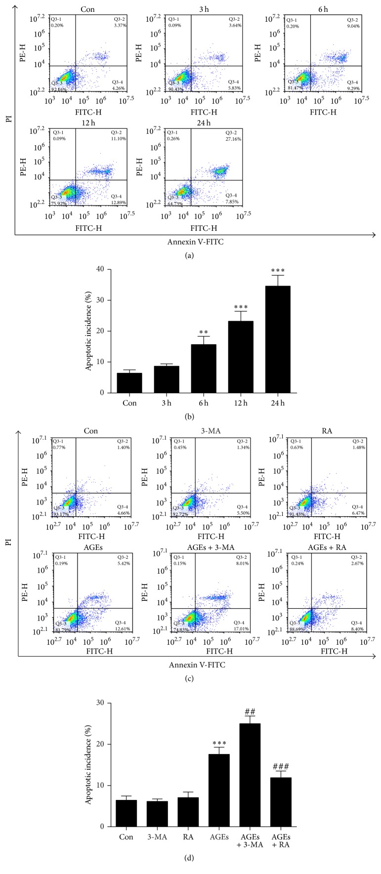
Figure 3.
Autophagy against AGE-induced apoptosis in chondrocytes. (a) Apoptosis in chondrocytes was measured by Annexin V/PI double-staining assay after treatment with 100 μg/mL AGEs for 0, 3, 6, 12, and 24 h. (b) Statistical analysis of flow cytometry results for apoptotic percentage of chondrocytes treated with AGEs. (c) Annexin V/PI double-staining assay for the apoptotic proportion of chondrocytes after pretreatment with 3-MA (5 mM) or RA (5 μM) for 1 h, followed by 100 μg/mL AGEs for 6 h. (d) The effect of autophagy on AGE-induced apoptotic rate. All results are the mean ± SD of three independent experiments. ∗∗p < 0.01 and ∗∗∗p < 0.001 versus the control group. ##p < 0.01 and ###p < 0.001 versus AGE-treated cells.

