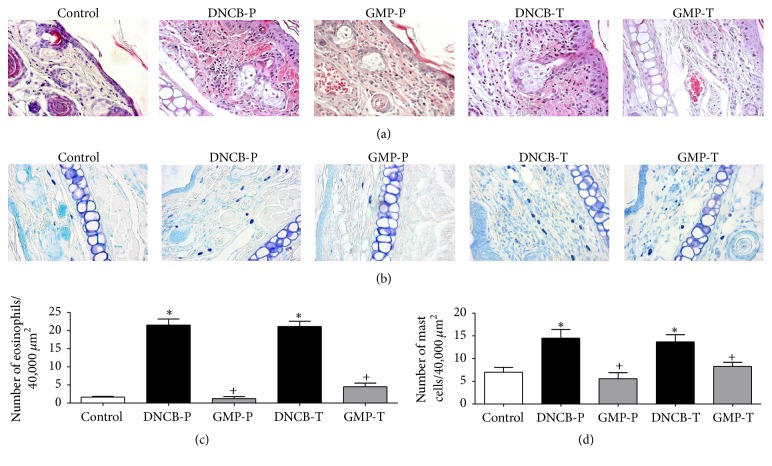
Figure 5.
Effect of GMP on inflammatory cell infiltration. Sections of right ears were stained with (a) hematoxylin and eosin to identify eosinophils and (b) blue toluidine for mast cells. Quantitative analysis of (c) eosinophils and (d) mast cells per 40,000 μm2 of dermis was developed with a microscope at magnification of 400x. Data are presented as mean ± SEM, N = 8. Control, not sensitized and water administered before AD-induction; DNCB-P, DNCB sensitized and water administered before AD-induction; GMP-P, DNCB sensitized and GMP administered before AD-induction; DNCB-T, DNCB sensitized and water administered after AD-induction; and GMP-T, DNCB sensitized and GMP administered after AD-induction; ∗p < 0.0001 versus control; +p < 0.0001 versus the respective DNCB without GMP administration.

