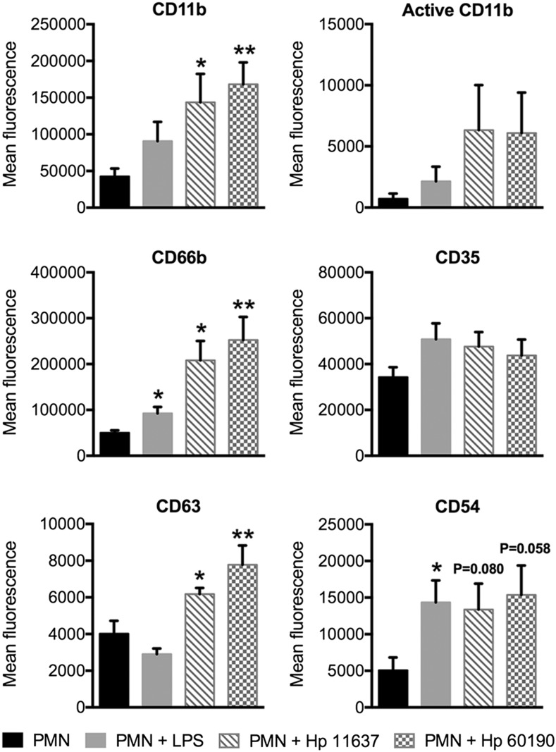
FIGURE 4.
H. pylori-infected neutrophils display additional surface markers indicative of subtype differentiation. Mean fluorescence of CD11b, active CD11b, CD66b, CD35, CD63, and CD54 was assessed by flow cytometry at 24 h. H. pylori-infected neutrophils and neutrophils treated with 10 ng/ml E. coli LPS were compared with untreated controls. n ≥ 4 donors. *p ≤ 0.05, **p ≤ 0.01.

