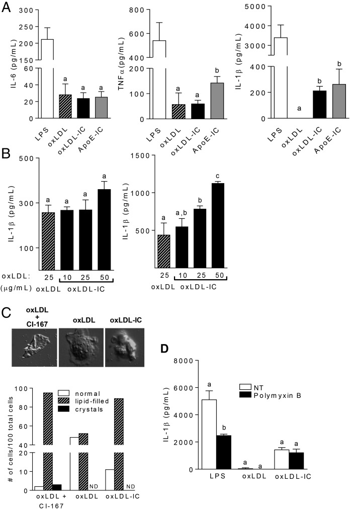
FIGURE 1.
oxLDL ICs prime the inflammasome. BMDCs were treated for 24 h with oxLDL or oxLDL ICs. (A) Cytokine levels in culture supernatants were measured by ELISA. Shown are representative experiments with n ≥3 biological and technical replicates. Unlike letters denote significance (p < 0.01) by Student t test, and error bars indicate SEM. (B) oxLDL ICs were tested for their ability to act as an activating (left panel) or priming (right panel) signal for the inflammasome. Briefly, BMDCs were treated for 3 h with 20 ng/ml LPS, followed by oxLDL or increasing concentrations of oxLDL ICs (based on oxLDL concentration) for an additional 3 h (left). For priming experiments (right panel), BMDCS were treated for 3 h with oxLDL or increasing concentrations of oxLDL ICs, followed by 5 mM ATP for 1 h. Culture supernatants were tested for IL-1β by ELISA. Shown is one representative of three experiments with three mice per experiment. Unlike letters denote significance (p < 0.05) by Student t test, and error bars represent SEM. (C) BMDCs were treated with oxLDL or oxLDL ICs for 3 h or with oxLDL in the presence of the ACAT inhibitor CLI-067 (positive control) for 24 h, and crystal formation was analyzed by polarizing light microscopy. Lipid-filled cells and crystal formation were quantified; representative images are depicted. Shown is one representative of two experiments. Original magnification ×1000. (D) BMDCs were treated with oxLDL ICs in the presence of polymyxin B. Shown is one representative of two experiments. IL-1β in culture supernatants was measured by ELISA. Unlike letters denote significance (p < 0.01) by one-way ANOVA with a Bonferroni posttest, and error bars represent SD.

