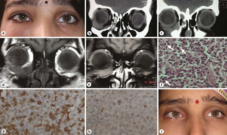
Fig. 1.
a Preoperative clinical photograph of the patient showing fullness of the right lower eyelid. b, c Computed tomography scan (coronal section) of the orbit showing a well-defined, hyperdense mass in the inferior orbit that appears to be arising from the orbital floor and medial wall anteriorly and extending toward the globe laterally and posteriorly. d, e Magnetic resonance imaging (coronal section) of the orbit showing a well-defined mass in the inferior orbit isointense to the muscle on T1-weighted images with good post-contrast enhancement and no mass in the PNS. f Microphotograph showing a cellular lesion with pleomorphic nuclei and numerous mitotic figures (arrows). HE. ×200. g, h Tumor cells showing positivity for immunohistochemical stains for CD3 and CD56, respectively (×400). i Clinical photograph of the patient at the last follow-up.

