Fig. 1.
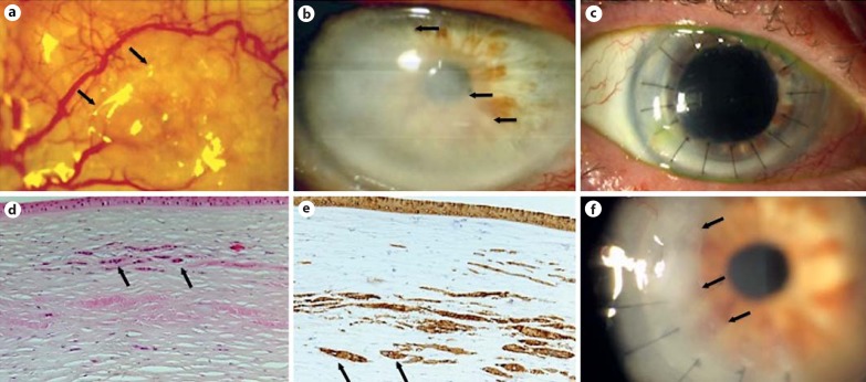
Case 1. a Slit-lamp photograph of OSSN recurrence demonstrating a conjunctival lesion at 9 o'clock, with a mildly elevated, gelatinous appearance and feeder vessels (arrows). b Slit-lamp photograph of the cornea demonstrating corneal haze originating from the cataract incision site that has crossed into the visual axis (arrows). c Slit-lamp photograph of a clear corneal graft after PK (postoperative day 63). d, e Histopathology of the corneal button shown in b, demonstrating midstromal infiltrative carcinoma with a normal overlying epithelium. Atypical cells that contain prominent nucleoli are present within the corneal stroma (black arrows) and stain positive for cytokeratin A1-A3 [HE. magnification ×200 (d); cytokeratin A1-A3, magnification ×200 (e)]. f Slit-lamp photograph of the corneal graft with recurrence of the midstromal haze (arrows) 1 year after grafting.

