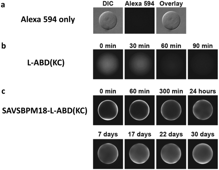
Figure 4. Spatial distribution of monomeric L-ABD(KC) and tetrameric SAVSBPM18-ABD(KC) on agarose beads (Sepharose 6B-CL) at different time points.

Time zero is the time point when the beads were mounted for fluorescence microscopy. (a) Alexa Fluor 594. The middle panel shows negligible retention of Alexa Fluor 594 dye alone on Sepharose 6B-CL under an epifluorescence microscopy. A differential interference contrast (DIC) filter was applied to visualize the presence of the agarose bead in the left panel. The right panel shows an overlay from both DIC and fluorescence images. (b) Alexa Fluor 594 labeled L-ABD(DC) and (c) Alexa Fluor 594 labeled SAVSBPM18-L-ABD(KC).
