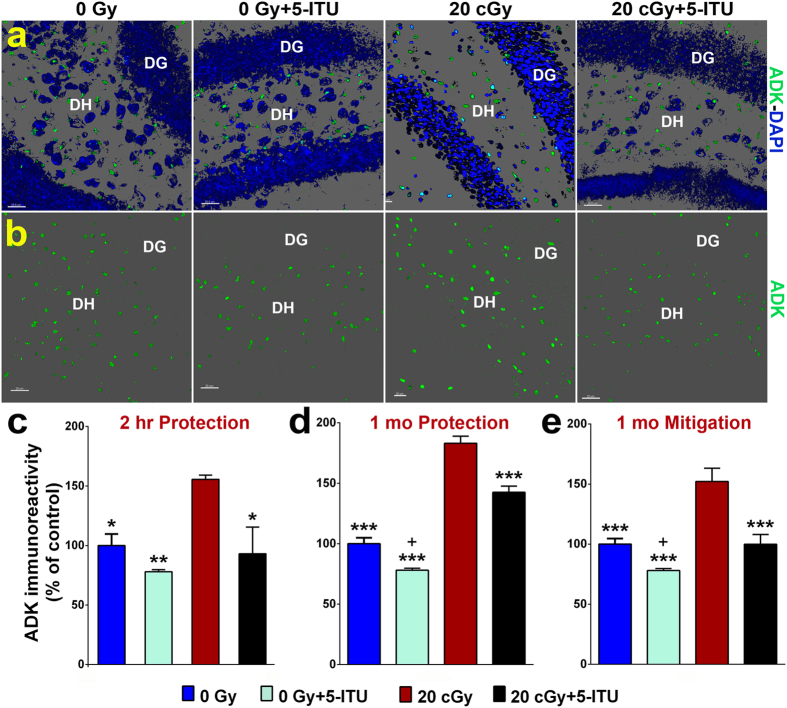
Figure 3. 5-ITU treatment protects against 28Si particle irradiation induced increased ADK immunoreactivity.
(a,b) Representative images illustrate that ADK protein levels are elevated 1 month post-irradiation (20cGy) that is reduced by 5-ITU treatment (ADK, green; DAPI nuclear counterstain, blue) in the hippocampal dentate hilus (DH) and dentate gyrus (DG). Deconvolution of images and quantification of ADK protein demonstrate that irradiation increased ADK at (c) 2 hrs post-irradiation and at (d,e) 1 month post-irradiation and that 5-ITU treatment protects against and mitigates this increase. Data are presented as mean ± SEM (N = 3–4 mice/group). P values are derived from ANOVA and Bonferroni’s multiple comparisons test (*P < 0.05, **P < 0.01, ***P < 0.001 for 0Gy, 0Gy + 5-ITU and 20cGy + 5-ITU as compared to 20cGy). +P < 0.01 for 0Gy as compared to 0Gy + 5-ITU. (a,b) Scale bars 20 μm.

