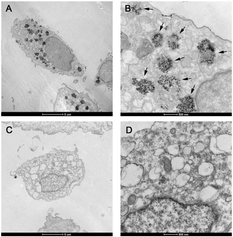
Figure 5. Transmission electron microscope for USPIO-labeled cells.
Canine ADSCs were treated with USPIO nanoparticles at iron concentrations of 50 μg/ml, Transmission electron microscopy indicated, compared with unlabeled cells, the USPIO labeled cells can also keep its inherent structure and morphology, and labeled ADSCs had iron nano-particles (black granular material) in the cytoplasm, as indicated by arrowheads. (A): USPIO labeled ADSCs, ×1650, scale bar: 5 μm; (B): USPIO labeled ADSCs, ×11000, scale bar: 0.5 μm; (C): unlabeled ADSCs, ×1650, scale bar: 5 μm; (D): labeled ADSCs, ×11000, scale bar: 0.5 μm.

