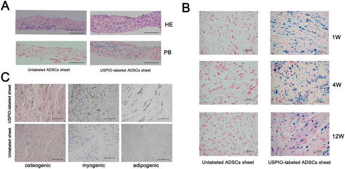
Figure 8. Histological analysis and differentiation of USPIO-labeled ADSCs sheet in vivo. 50 μg Fe/ml USPIO-labeled and unlabeled ADSCs continuously culture and form cell sheet with the stimulation of vitamin C.
(A): Histological analysis of USPIO-labeled cell sheet in vitro, HE represent HE stained, PB represent Prussian blue staining, scale bar: 100 μm. (B) Unlabeled (control) and 50 μg Fe/mL labeled ADSCs sheet were subcutaneously transplanted in the back of nude mice. Prussian blue staining for iron after 1, 4, and 12 weeks after transplantation, the blue-stained iron particles in the cytoplasm decrease gradually from week 1 to week 12, scale bar: 50 μm. (C) After 12 weeks of implanting, no spontaneous osteogenic, adipogenic and myogenic differentiation was observed in both USPIO-labeled and unlabeled ADSCs sheet, scale bar: 50 μm.

