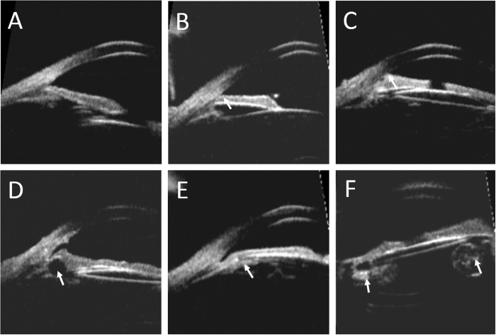
Figure 1. Abnormities in the configuration of the anterior segment in ultrasound biomicroscopy images.
(A) The normal angle structure of a phakic normal control. (B) High insertion of iris in the pediatric pseudophakia. The iris root was located more anteriorly than that of normal control. (C) Peripheral anterior synechia of iris. (D) Image of an iridociliary cyst causing localized angle narrowing. (E) An enlarged Soemmering’s ring causing angle narrowing. (F) Image of residual lens material in the capsular bag.

