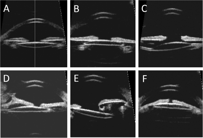
Figure 2. The intraocular lens (IOL) position in ultrasound biomicroscopy images.
(A) IOL implantation in the capsular bag with good position. (B) IOL implantation in the ciliary sulcus with good position. (C) IOL tilting due to asymmetric fixation with optic and one haptic in the bag while the other haptic in the sulcus. (D) IOL decentration and anterior synechia of iris. (E) IOL subluxation due to insufficient support of capsular bag. (F) IOL forward shifting and embedding into iridociliary tissue.

