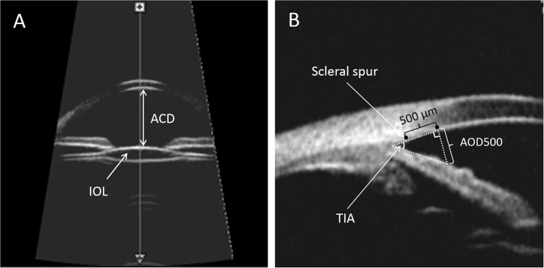
Figure 3. Ultrasound biomicroscopy (UBM) of the anterior segment of pediatric pseudophakia.
(A) UBM images of the anterior chamber and intraocular lens (IOL). Anterior chamber depth (ACD) was determined from the central inner corneal surface, perpendicular to the corneal surface to the most anteriorly visible part of the IOL. (B) Quantitative angle measurement. Angle-opening distance at 500 μm (AOD500) was measured on a line perpendicular to the trabecular meshwork at points 500 μm from the scleral spur. Trabecular-iris angle (TIA) was measured with the apex in the iris recess and the arms of the angle passing through a point on the trabecular meshwork 500 μm from the scleral spur and the point on the iris perpendicularly opposite.

