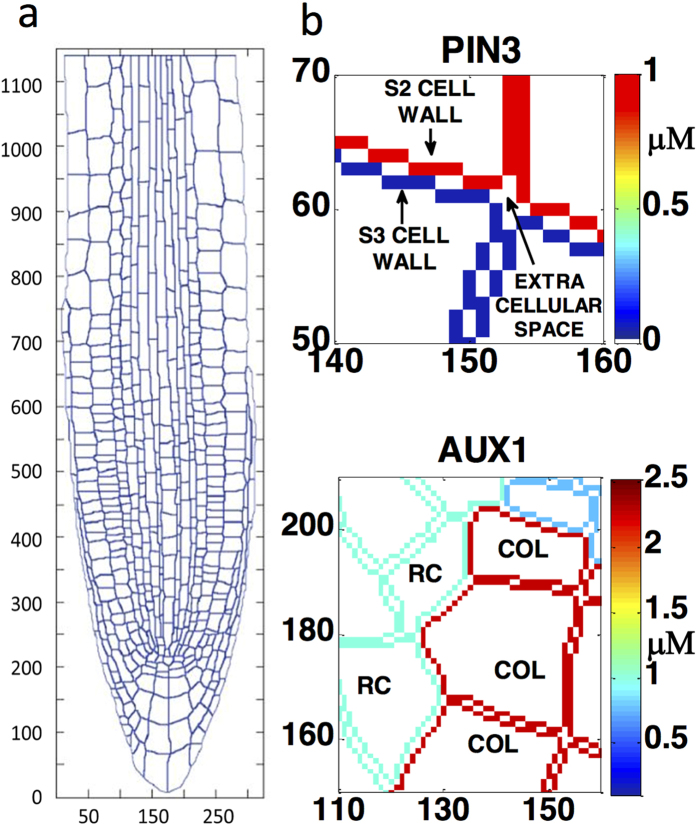
Figure 1. The model root with realistic cell geometry, a cytosolic space for each cell, a unique combined plasma membrane and cell wall entity for each cell, extracellular space, and auxin influx and efflux carrier localisation.
(a) A realistic root map showing the individual cells, based on confocal imaging13. (b) Localisation of efflux (PIN3) and influx (AUX1) carriers at the combined plasma membrane and cell wall entity of selected cells, with extra-cellular space between the cell walls of adjacent cells. (S2 and S3: columella tier 2 and 3 cells. COL: columella, RC: root cap). The details of the data-driven mechanistic model are included in the Supplementary Methods.

