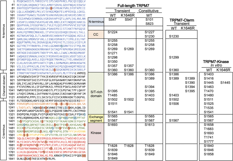
Figure 2. Identification of phosphorylated residues on TRPM7 by LC-MS/MS.
The diagram on the left depicts the amino acid sequence of mouse TRPM7 (UniProtKB: Q923J1). The cytosolic N-terminus, the coiled-coil domain, the S/T-rich domain, the exchange segment, and the alpha-kinase domain are colored in blue, orange, green, yellow and red respectively. Phosphorylated residues identified exclusively in the TRPM7-WT samples are highlighted with red circles. Sites also identified in the K1646R samples are shown in blue circles. The chart on the right summarizes identified phosphorylation residues from various TRPM7 constructs used in the study.

