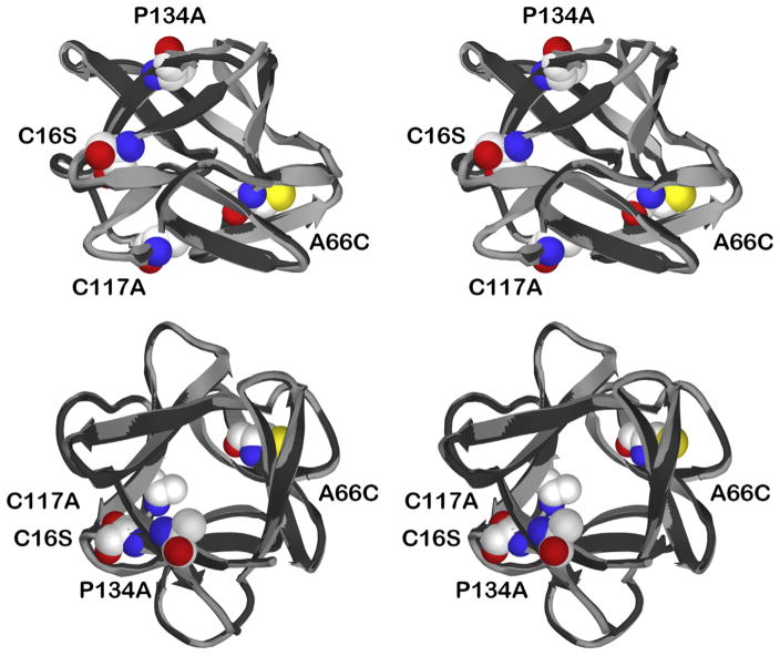
Figure 1.
Relaxed stereo ribbon diagram overlay of WT FGF-1 and C16S/A66C/C117A/P134A mutant X-ray structures. The C16S/A66C/C117A/P134A mutant structure (light gray) is overlaid onto the WT FGF-1 structure (black). The images include a “side” view (top panel) and a “top” view (parallel to the pseudo 3-fold axis of cyclic symmetry; bottom panel). Also shown are space-filling representations of the mutant residues Cys16Ser, Ala66Cys, Cys117Ala, and Pro134Ala.

