
Fig. 2.
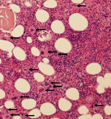
Bone marrow from patient with checkpoint inhibitor-induced ITP before rituximab treatment. H&E stained section of the bone marrow biopsy, 100 × magnification. The bone marrow is moderately hypercellular for age with trilineage hematopoiesis and increased megakaryocytes (black arrows) with a range of morphologies and mild clustering. These findings, coupled with the patient’s peripheral thrombocytopenia and elevated IPF, are compatible with a diagnosis of ITP