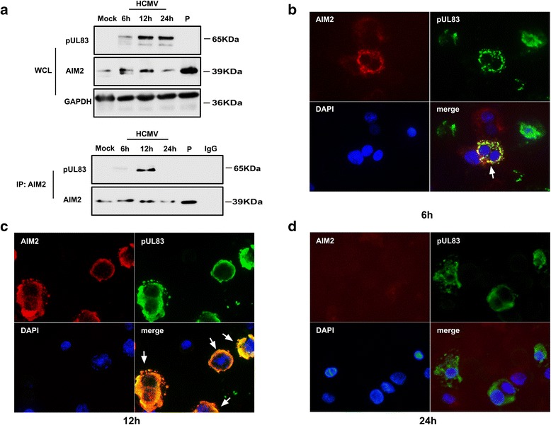
Fig. 3.

Detection of the pUL83/AIM2 interaction in HCMV-infected cells. a THP-1 cells were stimulated with PMA (100 ng/mL) to induce cellular differentiation. They were then mock-infected or infected with the HCMV AD169 strain for 6 h, 12 h, or 24 h, or transfected with poly(dA:dT). The cells were harvested and lysed and whole-cell lysates were immunoblotted using specific antibodies against pUL83 and AIM2, or immunoprecipitated with the anti-AIM2 antibody and then detected with anti-pUL83 and anti-AIM2 antibodies. b–d The infected cells were washed and fixed at the indicated time points. Specific antibodies against pUL83 and AIM2 were added and then conjugated with fluorescently tagged secondary antibodies. Cell nuclei were stained with DAPI. P: poly(dA:dT). WCL: whole cell lysates. Red, AIM2; green, pUL83; DAPI (blue), nuclei
