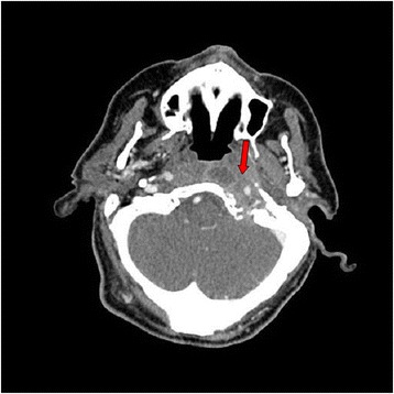
Fig. 2.

Cranial computed tomography scan shows a mass of soft tissue (arrow) surrounding the internal jugular vein and the carotid artery at the left jugular foramen with signs of bone erosion and destruction. The lesion extends medially and causes bone destruction of the left occipital condyle and the left side edge of the clivus and erosion of the posterior edge of the oval hole. In the petrous bone it extends to the middle ear and causes erosion of the anterior wall of the tympanic cavity. The mass goes along the petrous carotid reducing its caliber and causing bone destruction of the anterior edge of the carotid canal extending to the petrous apex
