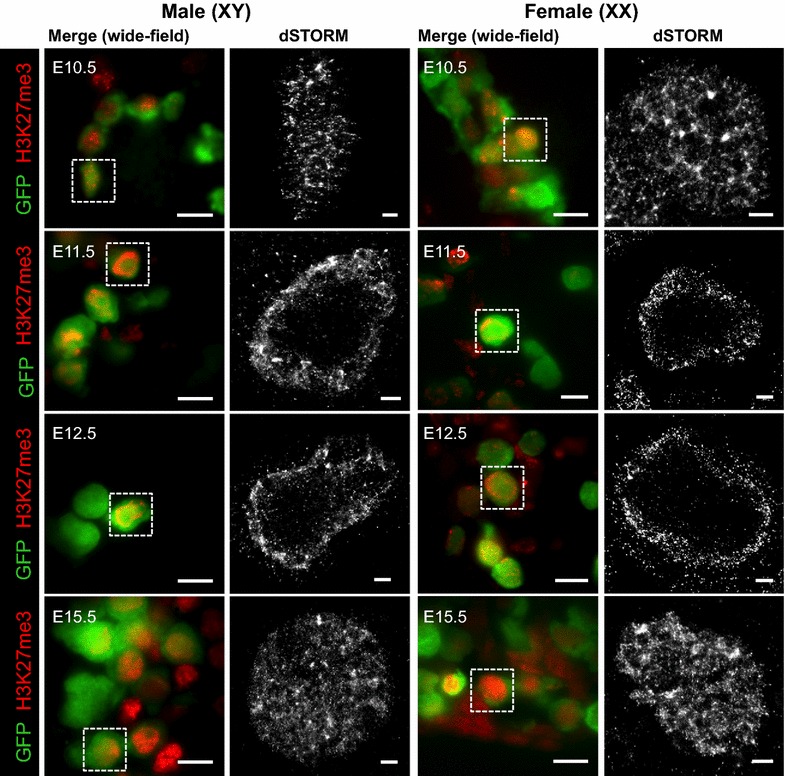
Fig. 6.

H3K27me3 is transiently relocated to the germ cell nuclear periphery in E11.5–E13.5 male and female germ cells. dSTORM super-resolution images in sections of XX and XY E10.5–E11.5 bipotential gonad and E12.5–E15.5 developing ovaries and testes. Left panels are merged wide-field (×160) images: eGFP marking germ cells (green), and H3K27me3 (red). White dotted boxes indicate super-resolved germ cell. 10 μm scale bars. Right panels dSTORM super-resolution of H3K27me3 antibody (grayscale). 1 μm scale bars. Representative images chosen from 3 to 8 super-resolved images in three biological replicates for each time point
