Fig. 1.

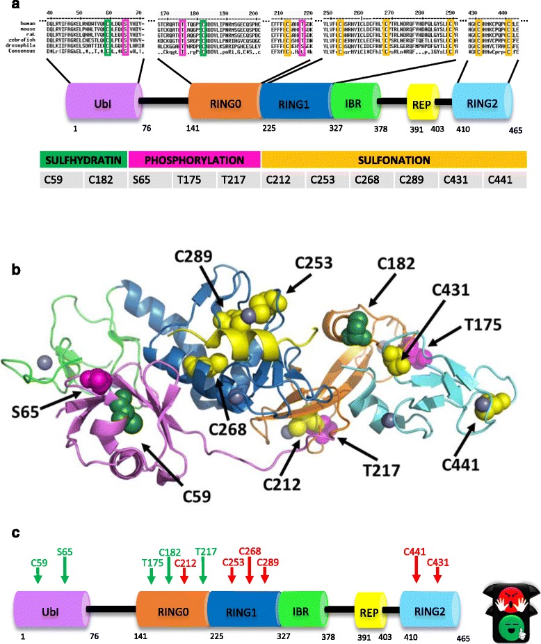
Mapping of Parkin Post translational modifications. a Domain architecture of Parkin protein and sequence alignment from different species. We differently highlighted the amino acids that are post-translationally modified (green: sulfhydration; pink: phosphorylation; yellow: sulfonation). Parkin consists of five domains: UBL, ubiquitin-like domain; RING, really interesting new gene; IBR, in between RING; REP, repressor element of Parkin. b Schematic representation of the full-length structure mapping the post-translational modifications onto the structure. c Primary structure and domains of Parkin mapping the activating (“green”) and inactivating (“red”) post-translational modifications
