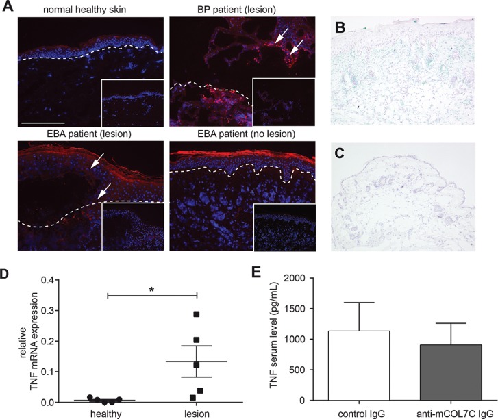
Figure 1.
Tissue expression of TNF was increased in experimental EBA. (A) Immunofluorescence staining of skin obtained from human BP and EBA patients and normal human skin. Red color indicates the presence of TNF only in lesional skin from EBA or BP patients. Inlay: isotype control staining. Images shown are representative of three specimens investigated per group. (B,C) Immunohistochemistry staining of ear skin obtained from (B) an anti-mCOL7C IgG-injected mouse and (C) a normal rabbit IgG-injected control mouse (scale bar: 400 μM). Blue color indicates the presence of TNF. (D) Ear samples from lesional skin (lesion, n = 5) and healthy areas (healthy, n = 5) after antibody transfers of anti-mCOL7C IgG were analyzed for mRNA levels of TNF by real-time PCR (n = 5/group, *p = 0.0382). (E) TNF concentration was measured by Bio-Plex in the serum of antibody transfer–induced EBA (anti-mCOL7C IgG, n = 7) and normal rabbit IgG–injected animals (control IgG, n = 4) 12 d after the first IgG injection. No significant difference was observed between the two groups.

